
Stanford University scientists have grown human brain tissue inside mice to create a new model for studying disorders such as cerebral palsy and frontotemporal dementia. But the real breakthrough is about giving those cells enough room to grow.

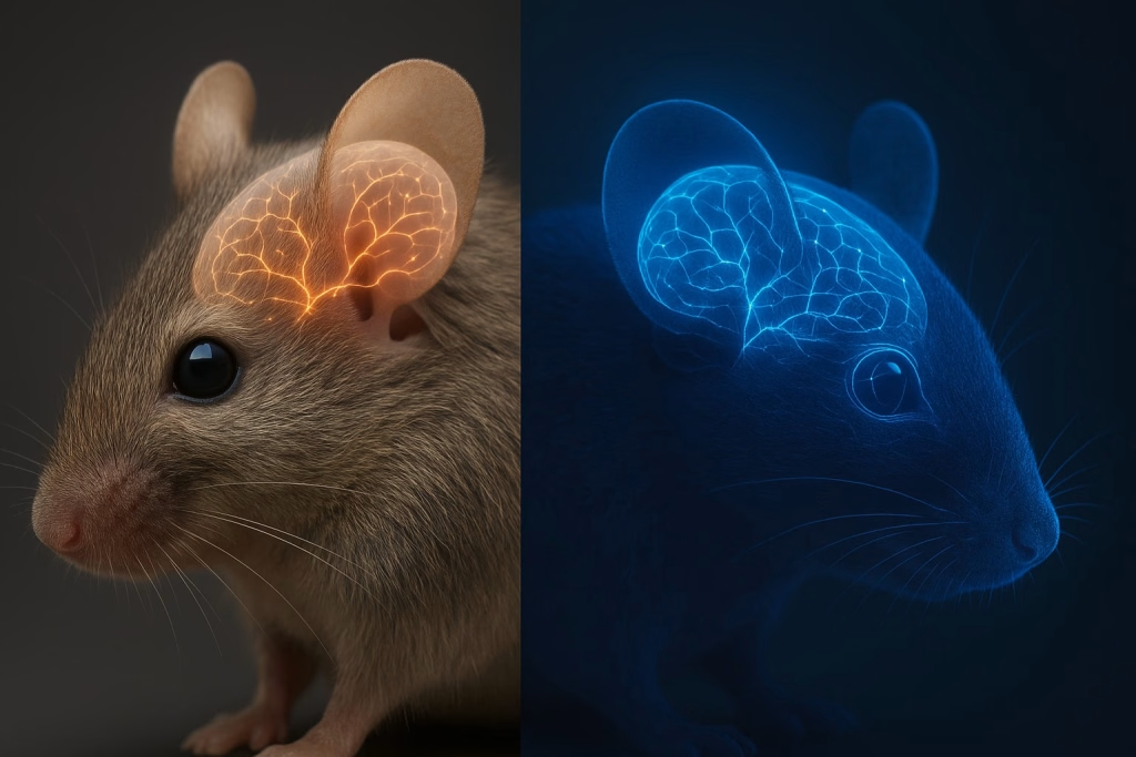
A team at Stanford University has created unusual mice in which lab-grown human brain tissue occupies more than 90% of the available cerebral cortex.
The research, published in Nature magazine on September 16, 2026, could give scientists a new way to study how human brain cells develop, respond to injury and behave in neurological disorders. The animals are called xenocortical mice.
It’s important to understand that scientists have not created a mouse with a human mind.
So, what exactly did Stanford scientists do?
The researchers first genetically engineered mice so that most of their cerebral cortex and hippocampus — key parts of the brain — did not develop. That left a large space where the mouse’s cortex would normally be.

Scientists then transplanted human cortical organoids into this space. These are tiny, three-dimensional pieces of human brain tissue grown from stem cells. Instead of competing with a normal mouse cortex, the human tissue had room to expand.
Within two to three months, it had grown nearly five times in volume, occupying more than 90% of the available cortical space. This is what makes the study different from earlier attempts to transplant human brain organoids into animals.
“For me, the real innovation is really removing the competition for space.”
That’s how developmental neuroscientist Giorgia Quadrato described the advancement to Nature magazine.


Why is this growth in mice brain cells important to humans?
Human brain cells mature much more slowly than mouse or rat brain cells. In earlier experiments, human organoids transplanted into rodents had to develop alongside the animal’s own rapidly growing brain tissue.
The Stanford team took a different approach: make space first, then let the human tissue grow.
The transplanted formed connections with the mouse’s nervous system, with human-derived nerve fibres extending as far as the spinal cord. That gives researchers something they cannot easily get from a dish of cells: human brain tissue developing inside a living nervous system.
Scientists found a rare type of human neuron

The experiment produced another surprise. The human tissue developed cells resembling von Economo neurons, specialised neurons associated with parts of the brain involved in social cognition. These cells are particularly interesting because they appear vulnerable in frontotemporal dementia (FTD).
Scientists had not previously been able to generate these neurons in standard laboratory cultures. Now, researchers can potentially study them while they are alive and functioning inside a biological system.
Stanford neuroscientist Sergiu Pașca, senior author of the study, said these cells are thought to be particularly vulnerable in frontotemporal dementia. That could eventually help researchers understand why some human neurons are damaged earlier or more severely than others in neurodegenerative disease.


The mice also revealed something about oxygen deprivation
The team then tested whether the model could reproduce a human-specific response to brain injury. They exposed the mice to low oxygen, or hypoxia.
The human brain tissue showed damage, and the animals developed problems with movement and coordination. The finding could be useful for studying brain injury associated with oxygen deprivation around birth — one area relevant to cerebral palsy research.
It is still an experimental model, not a treatment for cerebral palsy. But it gives researchers a way to study what happens to human-derived brain tissue during injury inside a living organism.
Did the mice become partly human?
No. The animals still have a mouse body and mouse nervous system. The human tissue is also not equivalent to a fully developed adult human cerebral cortex. Researchers found no evidence that the animals suddenly developed human-like intelligence or consciousness.
Why scientists are watching this development closely
Brain organoids have become an important tool for neuroscience because researchers can grow human-derived brain tissue without operating on a living human brain. But organoids grown in a laboratory dish have limitations.
They don’t have the complete environment of a living nervous system. The xenocortical mouse attempts to bridge that gap:
Human cells + living nervous system = a new platform for studying human brain disease.
Nature describes the work as the most extensive integration of human brain cells into an animal reported so far. Researchers could eventually use patient-derived cells to investigate particular genetic disorders or test potential therapies. But that future remains experimental.
The ethical question is even bigger
The more sophisticated these models become, the harder it is to separate the scientific question from the ethical one. A May 2026 report from the Nuffield Council on Bioethics examined the growing use of neural organoids and warned that increasingly complex models raise questions about animal welfare, possible changes in neural capacities and whether existing regulation is keeping pace with the science.
The Stanford researchers say their work involved consultation with ethicists and other experts. For now, the important question isn’t whether these mice are “half-human.”
It is how much human brain biology scientists can recreate in an animal — and where the ethical boundary should be as these models become more advanced.
The Innovators Jam ‘s take
The most interesting part of this experiment isn’t the phrase “half-human mouse.” It is the simple engineering idea behind it: remove the mouse cortex, create space, and let human brain tissue grow.
That has allowed scientists to see human neurons they could not previously study in a living system — and test how they respond to injury. If this can make brain-disease research more human-relevant, how far should scientists take it?
Mice engineered without most of their cerebral cortex and implanted with human cortical organoids.
More than 90% of the available cortical tissue was human-derived in the reported experiments.
Researchers believe the model could help investigate conditions including frontotemporal dementia, cerebral palsy and other neurological disorders.
No. They created a mouse model containing a large amount of transplanted human cortical tissue.
Found this article interesting? Subscribe to The Innovators Jam for news, views, and write-ups on innovation, science and research.
Follow us on Instagram @theinnovatorsjam, Youtube @InnovatorsJam, LinkedIn @ The Innovators Jam







We would love to hear from you. Leave your reply here.